





A visible narrowing or shadow across the middle section of the nose can sometimes develop after rhinoplasty. From the front, the border between the nasal bones and the upper lateral cartilages may form two diagonal lines resembling an upside-down letter “V”. This appearance is known as an inverted-V deformity.
Although the problem is often noticed because of its appearance, it may also indicate a loss of structural support in the middle nasal vault. Some patients consequently develop nasal obstruction, difficulty breathing during exercise or a sensation that the sides of the nose collapse inward when inhaling.
For patients considering revision rhinoplasty in Turkey, successful treatment requires more than hiding the visible lines. The surgeon must evaluate the nasal bones, upper lateral cartilages, septum, internal nasal valves and the overall width of the middle vault.
This guide explains why an inverted-V deformity develops, how it can affect breathing and which reconstructive techniques may be considered during revision surgery.
What Is an Inverted-V Deformity?
An inverted-V deformity is a visible contour irregularity that can occur where the nasal bones meet the upper lateral cartilages.
The upper third of the nasal bridge is primarily supported by bone. The middle third is formed by the upper lateral cartilages and the dorsal septum. Together, these structures create the middle nasal vault.
When the upper lateral cartilages lose support or separate from the nasal bones, they may move inward. The lower edges of the nasal bones then become more visible beneath the skin, creating the characteristic inverted-V outline.
The deformity may be:
- More visible on one side
- Symmetrical on both sides
- Mild and mainly cosmetic
- Associated with significant middle-vault narrowing
- Combined with internal nasal valve obstruction
- More noticeable in patients with thin skin
An inverted-V appearance may therefore represent both an aesthetic irregularity and a structural problem. Spreader grafts are commonly discussed in functional rhinoplasty because middle-vault complications can include inverted-V deformity, narrowing and internal nasal valve collapse.
What Is the Middle Nasal Vault?
The middle nasal vault is the central section of the nose between the nasal bones and the nasal tip.
Its main structural components include:
- The dorsal nasal septum
- The right upper lateral cartilage
- The left upper lateral cartilage
- Their relationship with the nasal bones
This area contributes to the smooth dorsal lines seen from the front and three-quarter views. It also contains the internal nasal valve region, one of the narrowest parts of the nasal airway.
A stable middle vault should maintain an appropriate relationship between the septum and the upper lateral cartilages. When this relationship is disrupted, the nose may look narrow, pinched, irregular or asymmetric.
The patient may also experience reduced airflow.
What Causes Middle Vault Collapse After Rhinoplasty?
Middle vault collapse most commonly develops when the supporting relationship between the nasal bones, upper lateral cartilages and septum has been weakened.
Several factors may contribute to the problem.
Dorsal Hump Removal Without Adequate Reconstruction
A dorsal hump may contain both bone and cartilage.
When the hump is removed, the upper lateral cartilages can lose their attachment to the dorsal septum. This creates what is sometimes described as an open-roof configuration.
The nasal bones may be repositioned, but the middle vault must also be stabilised. If the upper lateral cartilages are not adequately supported, they may move inward during healing.
This can narrow the bridge and create visible inverted-V lines.
Excessive Reduction of the Upper Lateral Cartilages
Aggressive removal of cartilage can weaken the middle third of the nose.
Once cartilage has been removed, the remaining structures may not be strong enough to resist the forces of scar contraction and breathing. The sidewalls can gradually move inward, particularly during deep inspiration.
Narrow Nasal Bones
Patients with short or narrow nasal bones may have a higher risk of visible middle-vault irregularities after reduction surgery.
When the nasal bones provide limited coverage over the upper lateral cartilages, even a small degree of inward movement may become visible.
Thin skin can make these structural borders more obvious.
Scar-Tissue Contraction
Scar tissue forms during every healing process.
After rhinoplasty, scar contraction may pull weakened upper lateral cartilages inward. This can make a mild structural weakness more noticeable over time.
In revision cases, scar tissue can also make the original anatomical planes difficult to identify.
Pre-Existing Nasal Asymmetry
A patient may already have:
- A deviated septum
- Unequal nasal bones
- Asymmetric upper lateral cartilages
- A crooked nasal bridge
- Previous nasal trauma
If these differences are not fully addressed during the first surgery, the middle vault may heal asymmetrically. One side can appear more collapsed or develop greater breathing restriction.
Trauma During Recovery
Significant pressure or injury during healing may affect the position of the nasal bones or cartilages.
Not every minor contact causes structural damage. However, a strong impact before the nose has stabilised may contribute to contour changes or asymmetry.
How Does an Inverted-V Deformity Look?
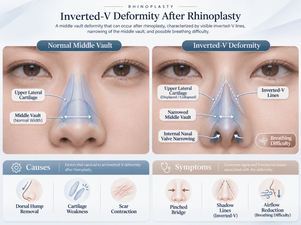
The deformity is usually assessed from several angles.
From the front, common signs include:
- Two diagonal shadow lines beneath the nasal bones
- A narrow or pinched middle third
- Interrupted dorsal aesthetic lines
- Unequal width on the right and left sides
- A bridge that looks wider above and narrower below
- A visible step between the bony and cartilaginous bridge
From the three-quarter view, the bridge may look irregular or indented. From the side, the deformity may be less obvious unless it is associated with a depression or an uneven dorsal line.
Lighting can change how visible the inverted-V outline appears. A clinical examination is therefore more reliable than a single photograph.
Is It Normal Swelling or an Inverted-V Deformity?
During the early stages of recovery, swelling can create shadows, temporary narrowing and asymmetry.
The nasal bridge may also look uneven after the cast is removed because swelling does not decrease at the same rate on both sides. These early changes do not necessarily represent permanent middle vault collapse.
Normal postoperative changes may:
- Vary throughout the day
- Improve gradually
- Look different under changing light
- Become less noticeable as swelling resolves
- Feel softer over time
A structural deformity is more likely to remain stable or become clearer as swelling decreases.
In most non-urgent cases, the final diagnosis should not be made too early. Revision rhinoplasty is commonly delayed until the tissues have healed sufficiently and the nasal structure has stabilised.
Patients with severe obstruction, progressive collapse, trauma, infection or skin changes should seek an earlier medical evaluation.
Can Inverted-V Deformity Cause Breathing Problems?
Yes. An inverted-V deformity can be associated with narrowing of the internal nasal valve.
The internal nasal valve is located between the septum and the caudal edge of the upper lateral cartilage. If the upper lateral cartilage moves inward, this space can become narrower.
Possible symptoms include:
- Difficulty breathing through one or both sides
- Increased obstruction during exercise
- Nasal sidewall collapse during inhalation
- Mouth breathing
- Difficulty sleeping with the mouth closed
- A sensation of insufficient airflow
- Improvement when the cheek is gently pulled sideways
- Temporary improvement with external nasal strips
However, breathing problems after nose surgery do not always come from the middle vault.
Other possible causes include:
- A deviated septum
- Turbinate enlargement
- Internal scar tissue
- External nasal valve collapse
- Tip collapse
- Nasal adhesions
- Allergic or inflammatory nasal disease
The American Society of Plastic Surgeons notes that revision patients may have internal valve collapse caused by deficiencies in the upper lateral cartilages and that cartilage grafts may be required to reopen this narrowed area.
What Is Nasal Valve Collapse After Rhinoplasty?
Nasal valve collapse after rhinoplasty occurs when an important part of the nasal airway becomes too narrow or lacks sufficient structural support.
Collapse may be static or dynamic.
Static Nasal Valve Narrowing
Static narrowing is present even when the patient is not taking a deep breath.
It may result from:
- Inward displacement of cartilage
- Septal deviation
- Scar tissue
- An excessively narrow middle vault
- Improper cartilage positioning
Dynamic Nasal Valve Collapse
Dynamic collapse becomes more noticeable during inhalation.
The negative pressure created while breathing pulls a weak sidewall inward. Patients may see the side of the nose move or feel the airway close when breathing deeply.
Some patients have both static narrowing and dynamic weakness. A spreader graft may widen the internal valve, but additional techniques may be needed when the lateral nasal wall itself is unstable.
How Is Inverted-V Deformity Diagnosed?
A detailed consultation should evaluate both appearance and nasal function.
The surgeon may examine:
- The width of the middle nasal vault
- Dorsal aesthetic lines
- The position of the upper lateral cartilages
- The nasal bones
- Septal alignment
- Internal valve width
- Sidewall movement during inhalation
- Scar-tissue formation
- Skin thickness
- Previous grafts
- Tip and external valve support
The examination may also include controlled support manoeuvres to determine whether opening a specific area improves airflow.
Patients should provide:
- Photographs from before the first surgery
- Recent photographs from different angles
- The date of every previous nasal operation
- Surgical reports when available
- Information about previous septal cartilage removal
- Details of nasal trauma
- A description of breathing symptoms
- Information about nasal sprays or allergy treatment
Online photographs can support an initial assessment, but they cannot show cartilage strength, internal scar tissue or dynamic valve collapse.
